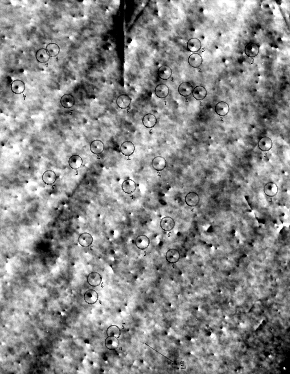
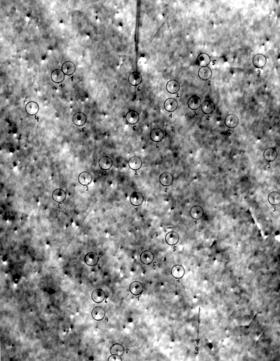
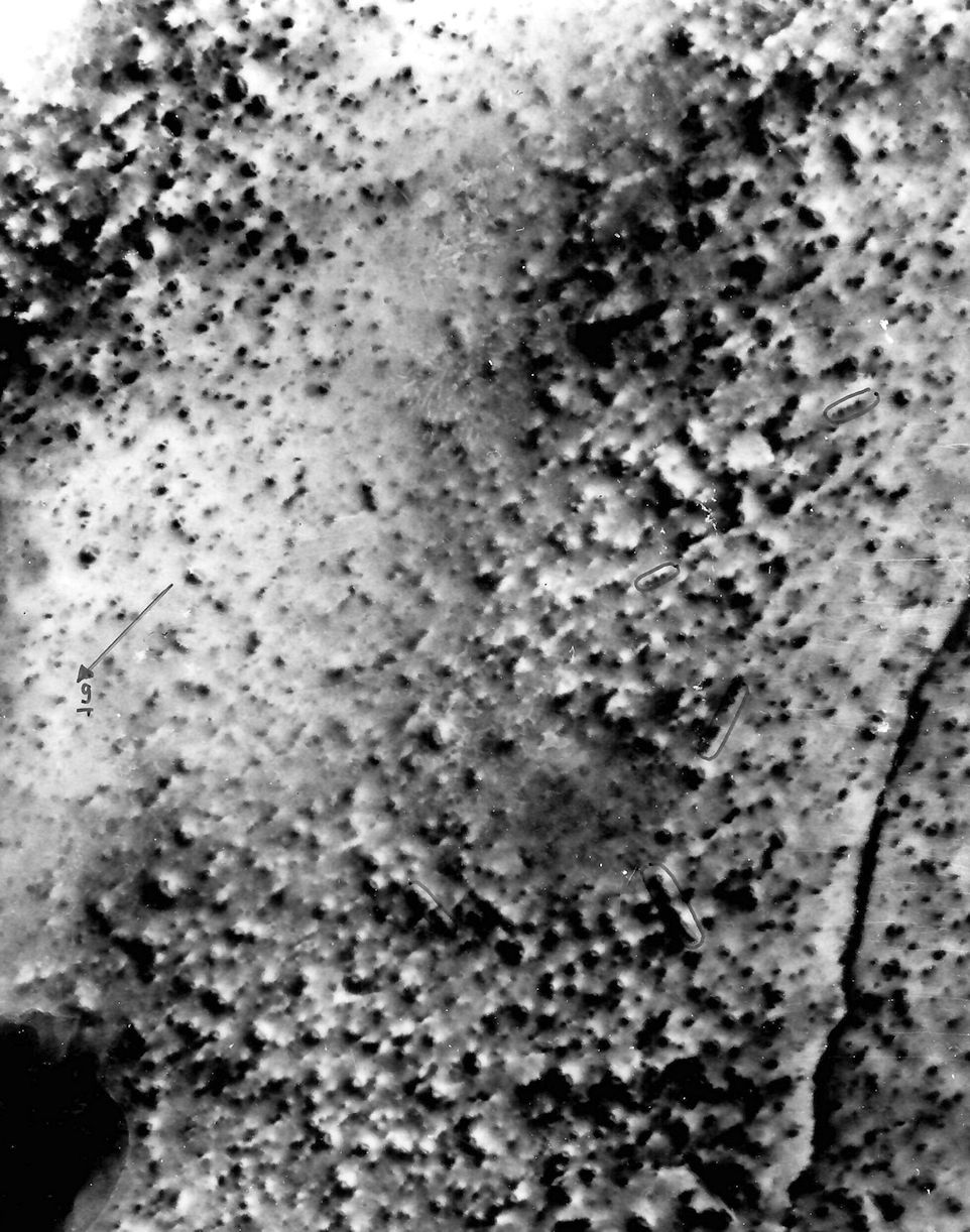
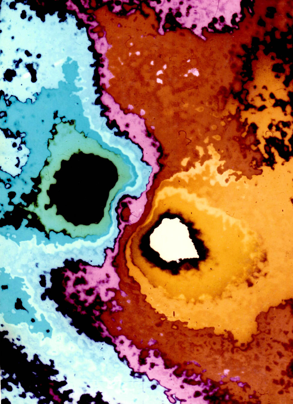
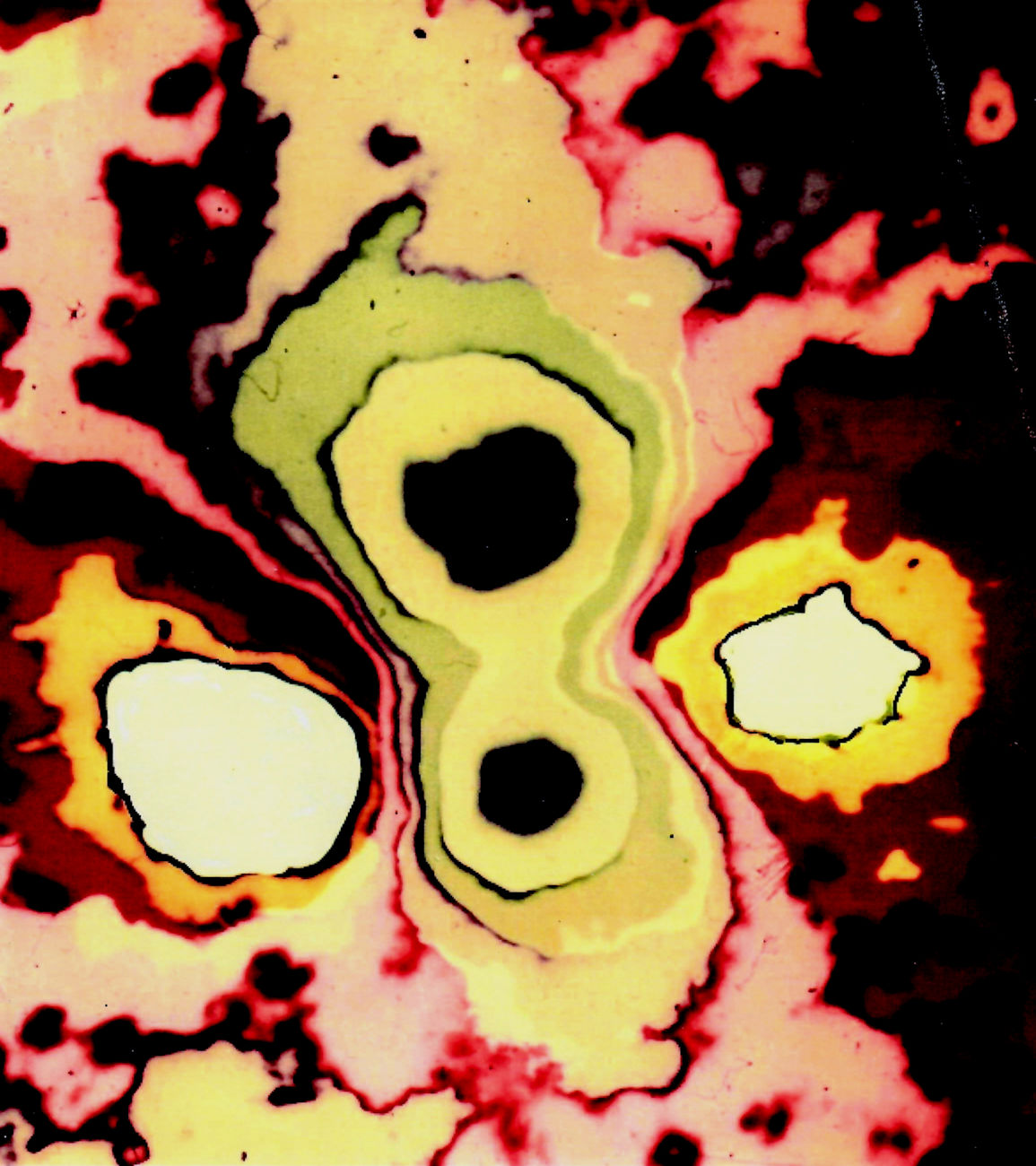
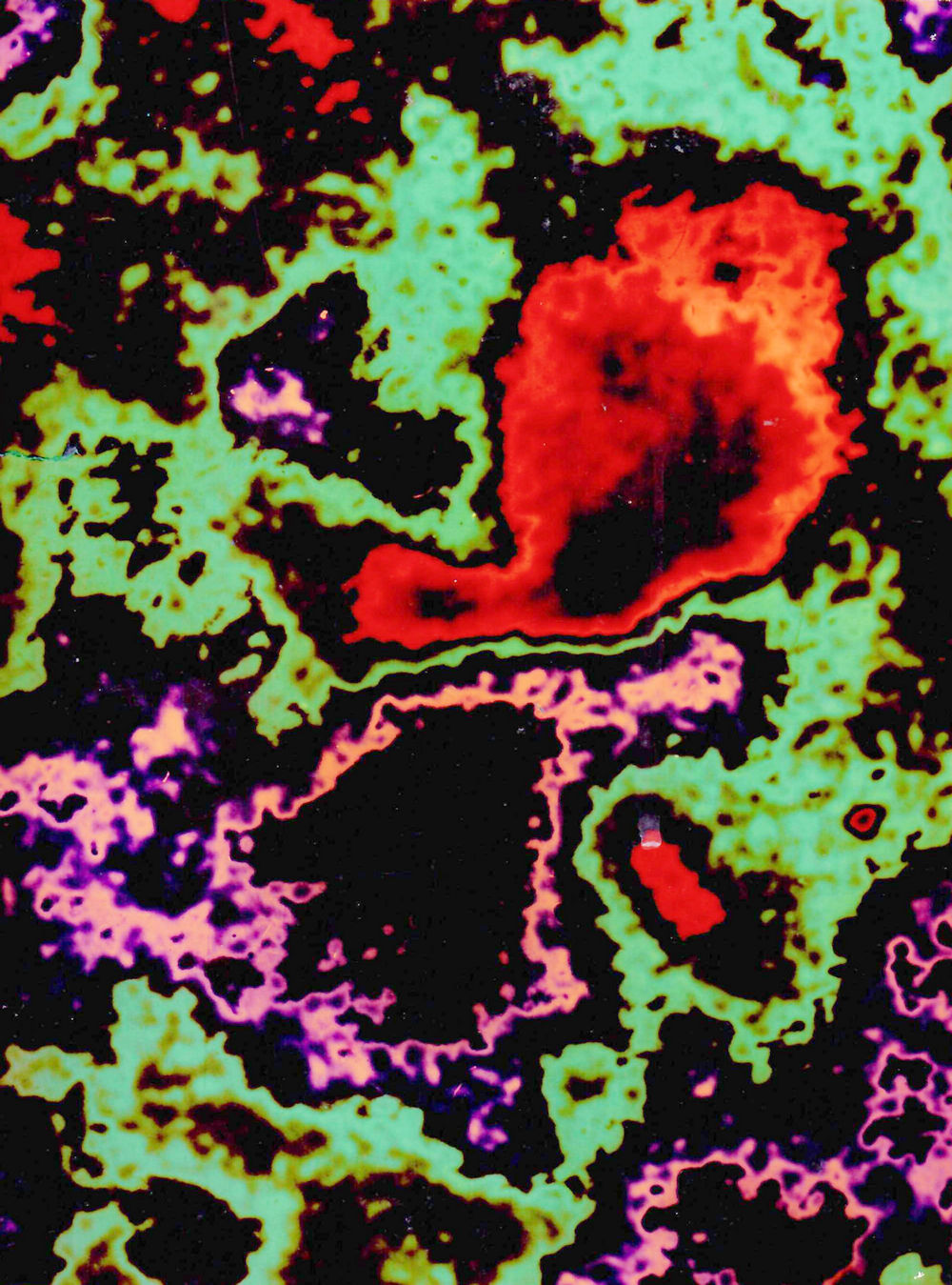
The picture expands to its full size when you click on it. Their size then is about what you would have produced on photographic paper in the dark room.
Like all the other ones, it results from a scan of the micrographs glued into my copy of my thesis paper. The negatives are long since gone.
And so it is. My kids will probably find it reassuring that I once was a sinner. But I did give the magnifications, after all, in the picture captions.
I also give you both the captions from my thesis and the ones from the publications.
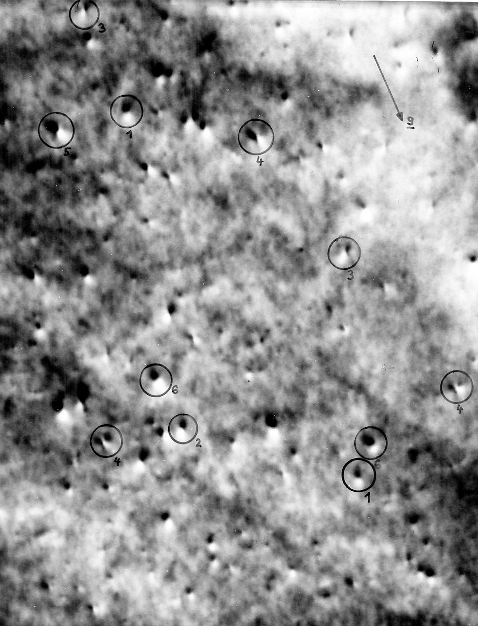 |
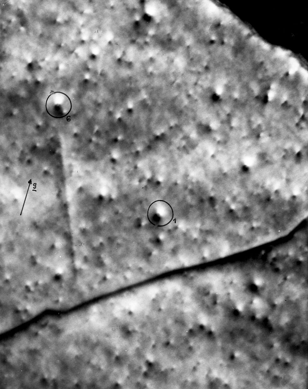
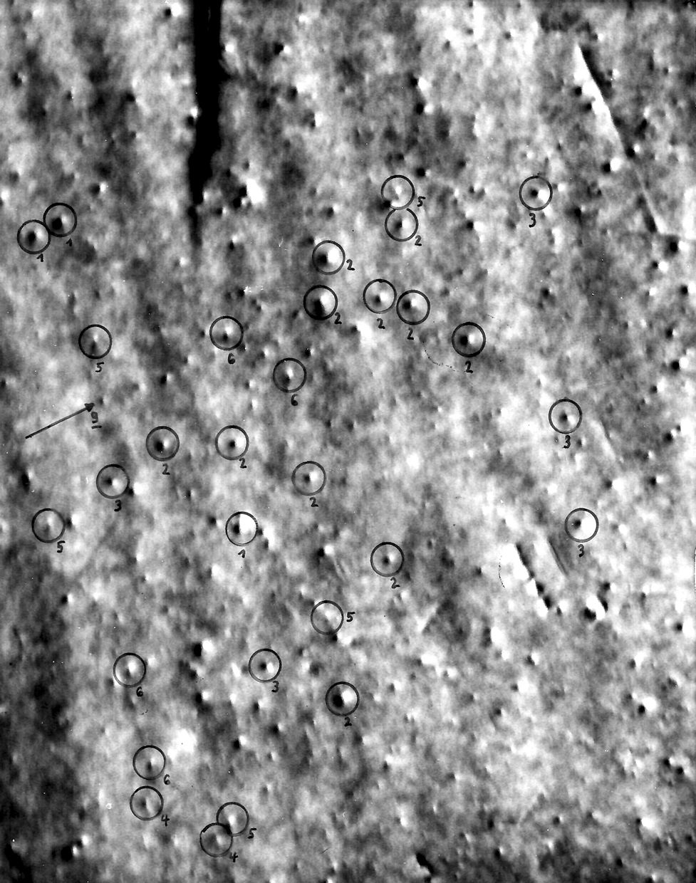
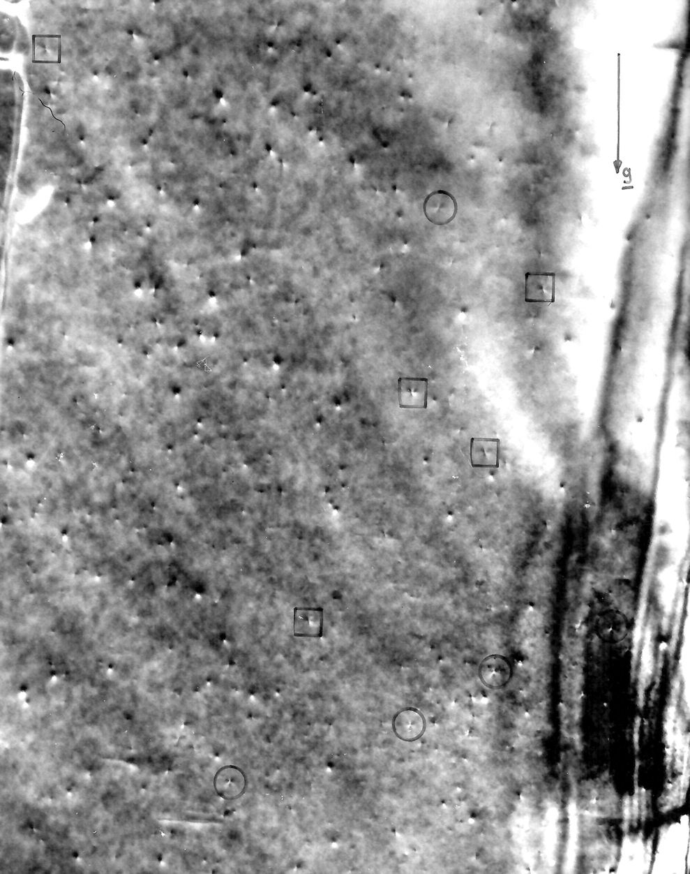
| Fig. 4.1 in Thesis. Original figure
caption:. Fig. 4.1. Typische Dunkelfeldaufnahme von S-W-Kontrasten bei Folienorientierungen nahe {0001} und g=11-20, v=500 000x, Dosis » 2,4 · 1011 cm2; 60 keV Au++ Ionen |
| Fig. 1 in paper 1 |
| Fig.l. D81·k field micrograph sbowing black-whitc coutrasts in Co. .Foil normal is near {OOOl}. Typical examples of thc A... B and C,. contrasts (sec text) arencircled. With respect to thc asteI'isks see Fig. 2 |
| Large size picture |
